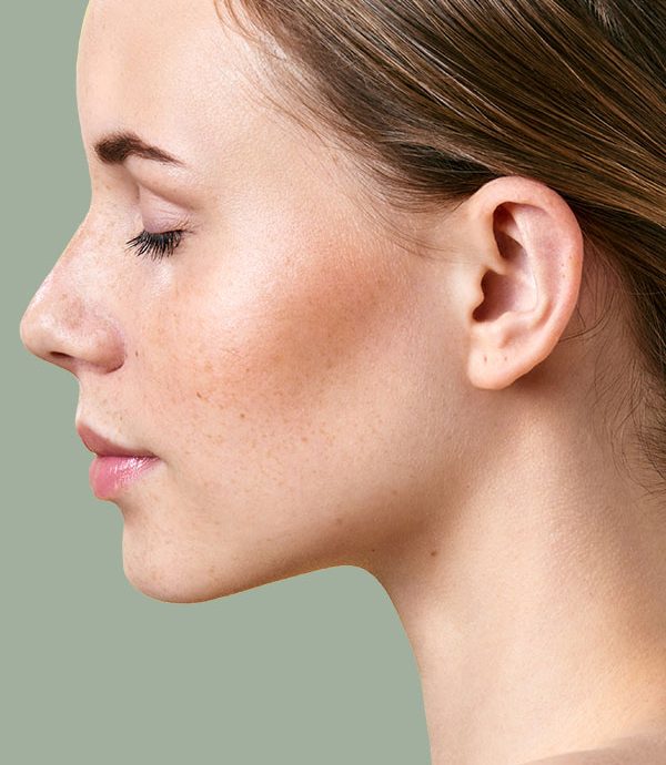
Otoplasty in Saarbrücken
Differences in the shape of the ear cartilage can cause the ear to protrude. Protruding auricles are the most common malformation of the ear. Over five percent of the population have protruding ears, commonly known as sail ears. The technical term for this is otapostasis (“prominent ear”). With protruding ears, the inner fold of the pinna is missing and the ear cavity is too large. The distance from the outer edge of the ear to the skull is greater. In ninety percent of cases, protruding ears occur on both sides. Protruding ears are often inherited. In around sixty percent of cases, there is a familial predisposition to protruding ears. Both men and women are equally affected by protruding ears. Hearing is not affected by the deformity.
What our patients say
Information at a glance

Operation duration
1 hour

Aftercare
6 weeks

Anesthesia
Twilight sleep

Thread tension
Self-dissolving threads

Hospitalization
Outpatient

Socially acceptable
after 2 weeks

Costs

Additions or alternatives

What is a pinnoplasty?
Ear pinning (otopexy) is used for the targeted correction of protruding ears. For a natural and balanced result, corrections of the ear cavity, the main fold of the ear and the earlobe are therefore combined. Lateral differences can be compensated to a certain extent. An optimal esthetic result can only be achieved by using the optimal technique. The techniques for correcting protruding ears have been continuously developed since Dieffenbach first described them in 1845. Over two hundred different techniques have now been described.
The techniques are named after the surgeons who gave them their names:
| Access at the back of the ear, incision of the cartilage in the area of the inner ear fold and the ear cavity as well as additional fixation sutures of the ear cavity (concho-mastoid sutures). This method is also known as the cut-and-sew technique. |
| Access at the back of the ear, incision of the cartilage in the area of the inner ear fold and the ear cavity as well as additional fixation sutures of the ear cavity (concho-mastoid sutures). This method is also known as the cut-and-sew technique. |
| Access at the back of the ear and suturing technique to restore the ear fold (mattress sutures). |
| Access at the back of the ear, incision of the cartilage in the area of the inner ear fold and the ear cavity as well as additional fixation sutures of the ear cavity (concho-mastoid sutures). This method is also known as the cut-and-sew technique. |
| Access at the back of the ear and fixation sutures of the ear cavity (concho-mastoid sutures). |
| Access at the back of the ear, incision of the cartilage in the area of the inner ear fold and the ear cavity as well as additional fixation sutures of the ear cavity (concho-mastoid sutures). This method is also known as the cut-and-sew technique. |
| Access to the front of the ear and thinning of the cartilage with a rasp to restore the ear fold. |
| Access at the back of the ear, incision of the cartilage in the area of the inner ear fold and the ear cavity as well as additional fixation sutures of the ear cavity (concho-mastoid sutures). This method is also known as the cut-and-sew technique. |
| Access to the back of the ear and thinning of the cartilage with a burr to restore the ear fold. |
| Access at the back of the ear, incision of the cartilage in the area of the inner ear fold and the ear cavity as well as additional fixation sutures of the ear cavity (concho-mastoid sutures). This method is also known as the cut-and-sew technique. |
| Access at the back of the ear, shaping of the ear fold with a transplanted cartilage strip of the auricle and fixation sutures of the ear cavity. |
| Access at the back of the ear, incision of the cartilage in the area of the inner ear fold and the ear cavity as well as additional fixation sutures of the ear cavity (concho-mastoid sutures). This method is also known as the cut-and-sew technique. |
| Restoration of the ear fold and fixation of the ear cavity without a skin incision (suture method). |
| Access at the back of the ear, incision of the cartilage in the area of the inner ear fold and the ear cavity as well as additional fixation sutures of the ear cavity (concho-mastoid sutures). This method is also known as the cut-and-sew technique. |
| Access at the front of the ear to restore the ear fold with a transplanted cartilage strip from the ribs. |
| Access at the back of the ear, incision of the cartilage in the area of the inner ear fold and the ear cavity as well as additional fixation sutures of the ear cavity (concho-mastoid sutures). This method is also known as the cut-and-sew technique. |
| The minimally invasive approach at the front of the ear for implanting U-shaped braces to restore the ear fold (Earfold®). |
Procedure for ear pinning surgery

Preparation
Before the ears are fitted, it is ensured several times that the best possible conditions are in place for an optimal result. You will be informed about the duration of the operation. You will be given medication to relax you and suppress the sensation of pain. The surgeon injects the local anesthetic around the ears using very fine cannulas. You hardly notice any of this.

The OP
The surgeon disinfects the ears and facial skin with an alcoholic solution. The hair and neck are carefully covered with sterile cloths. The surgeon checks the symmetry of the ears several times during the application of the ears. To do this, he carefully turns his head to the side alternately.
The planned new anthelical fold and incision are first marked on the skin with a thin skin pen. The surgeon works with magnifying glasses for precise execution.
The cartilage is exposed via an approximately four-centimeter incision at the back of the ear. The reconstructed ear fold is fixed with three to four fine sutures. The precise placement of the seams ensures a harmonious curve of the ear fold. Depending on the findings, part of the enlarged ear cavity is removed and the ear cavity is fixed with additional sutures (cavum pexy). These seams are placed with great sensitivity to achieve a natural shape. For a harmonious result, correction of the earlobe (lobule plastic) is not only necessary in rare cases. Where necessary, a narrow strip of excess skin is removed from the back of the ear. The skin is closed with fine sutures. A light pressure bandage stabilizes the new auricle shape and prevents the occurrence of bruising. The restoration of the natural ear fold takes about thirty minutes per side. A more comprehensive correction requires more time.

After the procedure
After the minor ear pinning procedure, you will be continuously monitored in the recovery room. Once you are fully awake, you can drink something and, if you can tolerate it well, eat a little. You will be supported by our nursing staff when you go to the toilet for the first time. Before leaving the practice, several appointments are made to check the wound. The surgeon will discuss with you again what you should bear in mind after the otopexy. You will be given a comprehensive written report and the surgeon’s personal telephone number. In an emergency, you can reach them 24 hours a day.


Six advantages of an ear correction
- Ear correction can significantly increase life satisfaction.
- Plastic surgery on the ears can prevent psychological stress.
- Parents often report an increase in their children’s self-confidence.
- After surgery, the ears are no longer covered by a haircut or by wearing a cap.
- Patients like to show off their ears and enjoy wearing ear jewelry in the months following the operation.
- The operation can be performed on an outpatient basis.
Before and after pictures of a pinnoplasty
Before/after photos on the subject of ears can be used in patient education, but may not be published online. We are therefore happy to show you before and after pictures of ears that have been operated on during the consultation. Of course, this is done in compliance with data protection regulations. The patients photographed have previously given their consent for this. You can find out more about this topic in the menu category “About us” under the menu item “Before and after pictures”.
Frequently asked questions
Protruding ears can have various causes. The most common cause is a combination of several factors:
- In most cases, the main fold of the pinna is too weak or missing completely.
- The ear cavity is sometimes too large and protrudes from the skull.
- The posterior ear muscle attaches too deeply to the ear cavity.
- Excess skin and a small muscle lead to a protruding earlobe (in about twenty percent).
The auricle is formed between the fifth and ninth week of pregnancy. At birth, the ear is very soft and easy to shape. By the age of three, the ears have already reached eighty-five percent of their adult size. In girls, the ears reach their final size one year earlier than in boys. The final width is reached at six to seven years and the final length at twelve to thirteen years. With increasing age, the elasticity and malleability of the ear cartilage steadily decrease.
In the newborn, ears that are blocked can be treated with plaster bandages. By the third month of life at the latest, however, the cartilage has lost its malleability. After the bandage is removed, the elastic ear cartilage returns to its original shape.
Tissue adhesive has been used in surgery since 1998. Tissue adhesives commonly used today consist of two naturally occurring proteins (fibrinogen and thrombin). The chemical skin adhesive cyanoacrylate is broken down by the body into formaldehyde and cyanoacetate. These metabolites can trigger an allergy and an inflammatory reaction in up to fourteen percent of patients 1 . After seven to ten days, the top layer of skin will naturally peel off. Even in newborns, where the cartilage is still soft, permanent success with skin glue is not to be expected. We strongly advise against the use of household adhesives. These adhesives are toxic and damage the skin.
The auricle is formed between the fifth and ninth week of pregnancy. At birth, the ear is very soft and easy to shape. By the age of three, the ears have already reached eighty-five percent of their adult size. In girls, the ears reach their final size one year earlier than in boys. The final width is reached at six to seven years and the final length at twelve to thirteen years. With increasing age, the elasticity and malleability of the ear cartilage steadily decrease.
The incidence of sail ears appears to be lower after birth than in the following years of life. Studies have therefore assumed that the ear cartilage is deformed by lying down. In the first few months of life, the ear cartilage is easy to shape. This property is utilized in the ear correction of newborns with silicone dressings. However, it is uncertain whether lying down has a significant influence and how high this influence is. What is certain is that in the majority of cases a predisposition leads to protruding ears. A varied sleeping position for babies also prevents head deformities caused by positioning. The baby’s head should be turned sometimes straight, sometimes to the left or right when sleeping in the supine position.
It is very important for infants to be accompanied and supported by their parents when putting on their ears. Otoplasty should be performed under general anesthesia. So that the children do not feel the injection on the back of the hand with the sleeping pill, the skin in this area is anesthetized with a special plaster half an hour before the procedure. After the ears have been positioned, the surgeon administers a long-acting local anesthetic (local anesthesia). This allows the children to wake up from the anesthetic without pain.
In small children, the ear cartilage is still soft. The cartilage can therefore be easily shaped into the desired form. The cartilage is then fixed with fine sutures. If necessary, an enlarged ear cavity is reduced in size and brought into the correct and desired position with sutures. Corrections of excess skin or the earlobe are performed at the end. The wound is closed with fine stitches. Careful bandaging of the ears is particularly important for children. On the one hand, the dressing must not be too tight, on the other hand, the children must not touch the wounds for reasons of hygiene.
Ear corrections must be carefully prepared. Tobacco smoking reduces blood flow to the tissue and impairs wound healing. Smoking has been shown to be associated with a higher risk of inflammation, even if it is stopped four weeks prior to ear pinning. The patient and the surgeon must carefully weigh up these risks in advance. Special precautions must be taken if you have concomitant illnesses such as high blood pressure or are taking certain medications.
Plastic and aesthetic surgery can be performed under light twilight sleep without any concerns. In adults, the ear cartilage is firmer. To bring the cartilage into the desired shape, the cartilage must be weakened or thinned out. Occasionally it is necessary to remove or transplant cartilage.
Adult patients should be collected after an operation.
- Avoid wearing glasses for the first ten days after the operation in order to keep the wound germ-free.
- Because of the medication you will be given during the procedure, you should strictly avoid driving on the day of the operation.
- Keeping the upper body elevated at thirty degrees and cooling for two to three days after the operation promotes decongestion and wound healing.
- You should make regular appointments to check your well-being and the results of the operation.
- A shaping bandage and an elastic bandage around the head are required for one week. You should wear a headband at night for the following five weeks.
- Avoid lying on your side at night for the first three weeks.
- Immediately before the wound check on the seventh postoperative day, you can take a normal shower and wash your hair.
- The skin sutures are removed ten to twelve days after the operation.
- Clean the newly formed ear fold behind the ear daily with a cotton swab after removing the skin threads. Then care for the skin with a neutral skin cream.
- Compliance with the hygiene rules ensures your surgical outcome to a high degree. Hands should be washed regularly with liquid soap for a period of thirty seconds. Avoid contact with animals.
- Avoid bending over. Lifting heavy loads, going to the sauna, swimming and other sporting activities should be avoided for 3 weeks and possibly even longer if there is still slight swelling.
- Avoid intense sunlight and extreme cold (skiing) for three to six months.
The thread method was described about twenty-five years ago. With the suture method or earplasty without incision, the surgical sutures are placed through the skin at the back of the ear. The knotted suture is placed through a small incision in the subcutaneous tissue. In order to weaken the restoring forces of the cartilage, the ear cartilage is cut through the skin with needle punctures (needle tear). The thread is sutured through all tissue layers: Skin, subcutaneous fatty tissue, cartilage and cartilaginous skin. The tying technique, the number of knots and the positioning of the sutures do not differ significantly from surgical techniques in which a skin incision is made. Bruising is not to be feared as the skin does not detach from the cartilage. Head bandages or headbands are therefore not used because the method is simple, painless and, unlike other surgical techniques, a scar-free procedure.
The suture technique without a skin incision has significant disadvantages. The ear cartilage cannot be thinned. Due to the restoring force of the cartilage, especially in adults, there is a greater risk that the auricle will return to its original shape. The stitches can protrude and promote inflammation. Excess cartilage or excess skin cannot be removed without a skin incision. A targeted and lasting correction of the auricle requires a skin incision.
In order to minimize the risk of inflammation, the wound is sterilely dressed after ear surgery. The wound is closed after about ten days. Until then, there is a risk of germs entering the wound and multiplying. It is therefore very important to observe hygiene measures. The dressing should not be changed independently for the first week. Hair washing is possible on the seventh day after the surgical procedure, immediately before the dressing change in the CenterPlast. The wound may only come into contact with water for a short time. Make sure that no soap residue remains on the scalp. The hair should be blow-dried with cold air. Wear the headband ordered for you until the dressing is changed in the CenterPlast.
The pinna reaches eighty-five percent of its adult size by the age of three. Protruding ears can therefore be corrected from the age of four. Children and parents should participate in the optimal follow-up treatment (visits to the doctor, hygiene rules, wearing a headband).
Children’s self-image is consolidated in the fifth year of life. Teasing at school affects self-esteem and self-confidence. The most recent studies on the ideal time for an otoplasty recommend surgery from the age of four. Otoplasty has been proven to improve children’s quality of life. Nevertheless, many patients undergo surgery in adulthood.
If patients are prone to proliferating scars, special precautions must be taken. In adulthood, secondary diseases, the use of blood-thinning medication and tobacco smoking can affect the outcome of the operation. In these cases, the patient and the plastic surgery specialist should discuss one of the most important questions: The risk-benefit ratio. This must be critically reviewed in this case.
Treatment is always associated with both opportunities and risks. It is therefore advisable to take out follow-up cost insurance before undergoing aesthetic procedures.
Scars heal slightly differently for each person. Children and adolescents have a strong immune system that tends to form pronounced, highly visible scars. Larger scars can form on the ears than on other parts of the body. People with dark skin color and people with a corresponding genetic predisposition can develop bulging or proliferating scars. Scarring can change the shape of the pinna and lead to asymmetries. Slight side differences are normal for everyone. Correcting asymmetries is therefore only worthwhile if they are really noticeable.
The restoring force of the cartilage can lead to the regression of the simulated inner ear fold (anthelix) after some time. Scarring can lead to excessive emphasis of the inner ear fold (anthelix). When looking from the front, the inner ear fold (anthelix) covers the outer ear fold (helix). In this case, the fold of the outer ear recedes into the background, hence the technical term “hidden helix”. Various causes can lead to the auricle being tighter in the middle than the upper edge or the earlobe. This unsatisfactory form is also known as the “telephone earpiece”.
If the cartilage has been shaped using an incision technique, tiny edges and kinks may become visible after the swelling has subsided. Unevenness of the auricle can be particularly noticeable if the skin is thin.
Suture granulomas or fistulas may form in the event of intolerance to surgical sutures. Inflammation of the skin or cartilage may require surgery and antibiotic treatment. In case of inflammation, an aesthetically unfavorable result and a conspicuous scar must be expected.
A feeling of numbness or hypersensitivity of the auricle usually disappears by itself after a certain time.
If you would like to find out the price of a specific ear correction operation, we recommend that you use our price calculator or arrange a consultation.
Health insurance will only cover the costs of ear pinning for minors if a psychologist’s or psychiatrist’s report shows that they are suffering a great deal from teasing. You can find further information on the topic of cost coverage by health insurance companies in the menu category “Costs” under the item “Obligation of health insurance companies to provide benefits”.

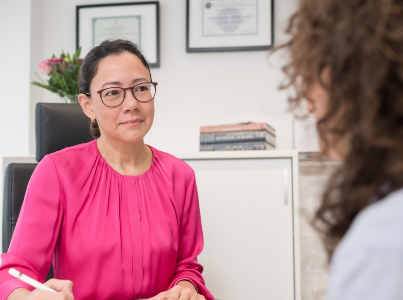
AUTHOR
Dr. Adelana Santos Stahl
Our aim is to offer optimal, discreet and precise treatment based on our extensive expertise in the field of plastic surgery.
Dr. Adelana Santos Stahl has a unique international perspective with a female view of plastic surgery. Your individual and detailed approach is the key to the beautiful and natural results. Having trained in Brazil, one of the largest and best-known countries for aesthetic and reconstructive plastic surgery, she understands her patients’ desire to look and feel their best.
She completed her medical studies and training as a specialist in plastic and aesthetic surgery in Brazil. In 2009, she also successfully passed the German equivalence examinations for the state medical examination.
Two years later, in 2011, she received the German and in 2012 the EU specialist certification (EBOPRAS) for plastic surgery. From 2009 to 2013, she deepened her knowledge of aesthetic and reconstructive facial surgery with world-renowned representatives of plastic surgery such as Professor Gubisch at the Marienhospital and Madame Firmin in Paris.
A VDÄPC Fellowship (continuing scholarship for graduate students) in Switzerland, France and the USA has further enriched her professional experience. Dr. Santos Stahl is active in various renowned professional associations. In addition to the DGPRÄC and DGBT, she is also a member of the Brazilian Society of Plastic Surgery – SBCP.
She is also the author of several scientific articles and, together with her husband, is dedicated to research and clinical studies in the field of plastic surgery.
She has been based in Saarbrücken since 2019.
- Nigro LC, et al., 2020, Plast Reconstr Surg ↩︎
You might also be interested in


Personal advice
We take time for you and offer you customized advice and treatment for your individual result.


